
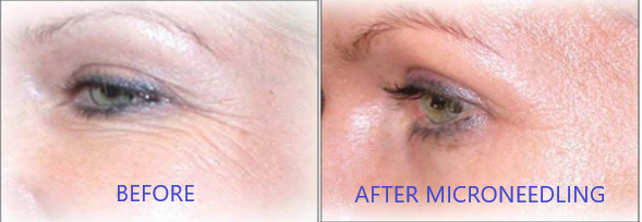
It is a widely accepted tenet of aesthetic medicine that, in order to stimulate new collagen formation in the skin, we have to injure the skin. The injury turns on healing mechanisms, including stimulating the cells that make scar tissue to make collagen.
For many years, the focus was on using whatever modality (laser, radiofrequency, ultrasound, microneedling) to create enough of an injury to get new collagen formation (and hence skin tightening) without getting ugly scars.
The attitude was “No pain/no gain” and people tolerated some very painful treatments.
History of microneedling
Horst Liebl was the original inventor of the Dermaroller and other microneedling devices. He also researched and identified the true mechanism by which microneedling achieves its amazing results.
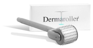 The Dermaroller, a short cylinder studded with almost 200 needles on an attached handle, was invented in 2000. It was originally intended as a method of creating channels in the skin through which therapeutic solutions could be infused.
However, the Dermaroller was found to produce effects such as lifting depressed scars, flattening raised scars and improving pigmentation, and these effects could not be ascribed to the solutions being applied to skin.
When they looked at skin biopsies of treated skin, they found that new collagen was forming at a depth of 0.5 to 0.6mm. To give some context, facial skin is about 1.5mm thick.
Researchers interpreted this depth of collagen formation as indicating that microneedling worked from the bottom up. Thus longer needles were predicted to give better results. No pain/no gain.
-

- Screenshot from Kim Kardashian’s video from 10 years ago, showing her microneedling treatment, illustrating the no pain, no gain attitude.
-

- Screenshot of Kim Kardashian’s Instagram post 10 years ago showing how she looked after her MN treatment
If you ever saw an early video of Kim Kardashian getting a microneedling procedure, her face looked like a slab of raw, bloody meat. No pain/no gain, indeed.
However, what researchers overlooked was that microneedling to a depth of 0.6mm or greater, resulted in the formation of type I collagen scar tissue, giving the skin an altered stiffer texture. Microneedling to depths less than 0.6mm resulted in a more youthful architecture of the skin with more type III collagen, elastin, thicker epidermis, and other youthful components such as proteoglycans and glycosaminoglycans, giving the skin a more youthful texture.
It turns out that microneedling ideally stimulates the basal layer of the skin, which is at a depth of 0.15mm. The benefits of microneedling spread downwards from the basal layer, rather working from the bottom up so longer needles do not improve results. In fact, longer needles can damage structures under the skin, not to mention make the treatment so painful that anaesthesia is required.
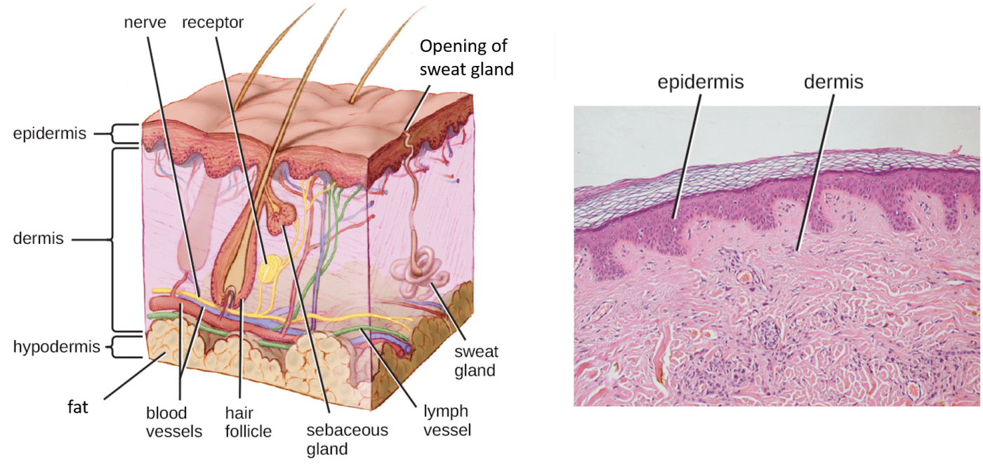
Left, a diagram of the full thickness of skin. The epidermis is the thin darker layer on top. The basal layer is the thin pinker line on the lower side of the epidermis. Right, human skin viewed under microscope. The epidermis is the dark pink layer and the basal layer is on the undulating lower margin of the epidermis. Note how the lighter pink dermis is full of structures. Those are cells, blood vessels, bands of collagen and so on.
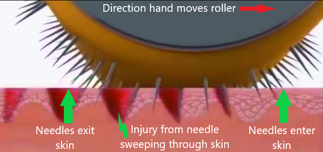
One of the drawbacks of the Dermaroller was that it gave a painful treatment. Here at Advanced Rejuvenation, we never offered the Dermaroller as a face treatment because we could never make it comfortable. It turns out that the act of rolling the Dermaroller makes the needles move through an arc in the skin, which is very painful.
The ACS Pen, invented by Horst Liebl, uses very fine needles in a straight line. The pen is held perpendicular to the skin so the needles enter and exit vertically. Our staff are trained to move the pen at the correct speed timed as the needles go up and down, so there is no injury outside of each precise pricking channel.
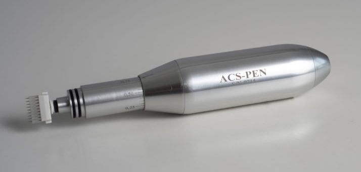
ACS Pen microneedling tool . Note the sterile cartridge of very fine needles on the left end.
Fun fact: microneedling pens were first based on tattooing equipment.
Detailed explanation of what’s happening at the molecular level (optional)
Our bodies can create battery-like effects at a cellular level by separating negatively charged ions from positively charged ones. In the case of skin, the surface of the skin has a negative charge and within the skin itself, the liquid between the cells (the interstitium) has a positive charge, and inside the cells of the skin exists a negative charge. When a difference in charge exists, it is called an electrical potential. The skin is used to keeping these electrical potentials in balance.
Keep in mind, we have 2 separate electrical potentials here: (1) between the skin surface and the interstitium (also called the extracellular space), and (2) between the interstitium and the inside of the skin cells (also called the intracellular space).
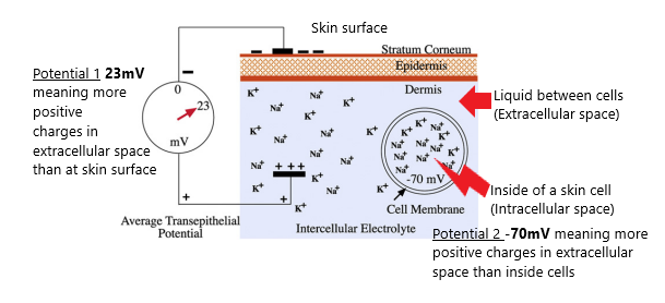
At rest, skin has differences in positively and negatively charged ions, creating electrical potentials, in 2 places: between skin surface and the extracellular space, and between inside skin cells and the extracellular space
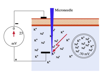
At the moment of microneedling, the electrical potential between skin and extracellular space collapses to zero
The initial pricking of the skin, either by injuring the skin or due to the electrical conductivity of the steel needles, causes the difference in charge between the surface and the interstitium to collapse. This change in electromagnetic field around the needle prick causes existing red and white blood cells to migrate to the pricking channel and close it up. Microneedling pricking channels seal up in less than 20 minutes.
The white blood cells work by releasing growth factors so this is one of the mechanisms for making more collagen in response to microneedling.
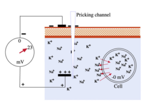
Immediately after the needle exits skin, the skin re-establishes the electrical potential to 23mV by pumping ions out of the cells, thus changing the electrical potential between the intracellular and extracellular spaces
The collapse of the first electrical potential also throws the 2nd electrical potential out of balance. Skin cells quickly work to reestablish the electrical potential so they turn on ion pumps to kick positively charged ions out of cells so that there are more positive ions in the fluid around the cells (extracellular space) than inside the cells (intracellular space).
They actually generate a higher electrical potential (meaning a higher concentration of positive ions outside the cells) and it is this higher-than-normal electromagnetic field that changes which genes are active in the stem cells of the skin. Undifferentiated stem cells switch over to a much more active form called progenitor stem cells.
Progenitor stem cells produce the various differentiated cells of the skin (such as keratinocytes, melanocytes and fibroblasts). These newly minted cells help thicken up the epidermis, making it more youthful, as well as stimulating the production of collagen, elastin, proteoglycans and glycosaminoglycans in the dermis.
Proteoglycans and glycosaminoglycans act as the stuffing in the skin, making it plumper and softer, akin to how better stuffing in a chair’s upholstery makes the chair more comfortable.
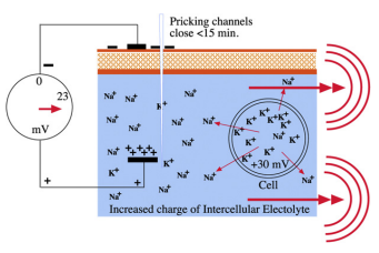
After skin cells pumped out Na+ ions, the electrical charge inside the cells has risen from -70 to +30mV, an electromagnetic field that causes stem cells to change into progenitor stem cells that rapidly differentiate into the various types of skin cells
The effects of altered gene expression start to become apparent within days and can continue to improve skin quality for months.
The ideal distance between pricking channels is at least 1mm apart. This is based on the distance that the newly-produced skin cells are found after microneedling, about 1mm away from the pricking channel.
I’ve gone into so much detail about the exact mechanism of how microneedling improves skin to illustrate why you don’t need a visible injury to get results.
Summary of why microneedling works
The equipment that we use, the ACS Pen, uses such fine needles that it is very unlikely that the needles even puncture or damage skin cells during the procedure but rather push cells out of the way to create the pricking channels.
The act of pricking the skin with these fine needles sets up tiny electromagnetic fields that stimulate migration of healing cells to the area of the pricking channel, as well as activating stem cells in the basal layer of the skin to differentiate into all of the cell types found in healthy youthful skin. The new cells migrating into the epidermis make it thicker leading to a more youthful skin appearance.
The healing cells and the cells generated by stem cells release cellular messengers like growth factors and cytokines, that will, in turn, stimulate the production of molecules that plump up and hydrate the dermis.
Microneedling does not have to be deep, because the target layer is only 0.15mm below the surface.
Microneedling does not give better results if we create more punctures by doing more passes, because the ideal spacing for each needle prick is 1 – 2 mm apart.

This is me, Dr Rose Jeans, immediately after a microneedling treatment
Thus microneedling does not have to be painful to give beautiful results. As you can see from the photo of me, immediately after a MN treatment, I’m only slightly pink with no other signs of injury.
Here at Advanced Rejuvenation, we boost the results of microneedling by applying growth factors or exosomes to the microneedled skin. I just had my first exosome treatment in this photo. We’re amplifying what is happening at the molecular level in the skin so that you will make more collagen and more new skin cells and get more rejuvenation of your skin.
Thus you will get lots of gain with minimal or no pain.